Advanced imaging technologies allow your veterinarian to visualize your pet’s internal structures and processes, identify hidden disease or abnormalities, collect diagnostic samples, and perform interventional treatments that provide life-saving or life-extending results. Central Kentucky Veterinary Center is proud to provide a suite of state-of-the-art imaging services that include digital radiography (i.e., X-ray), ultrasound, thermography, and computed tomography (CT).
When would imaging be recommended for my pet?
Advanced imaging is an invaluable and versatile diagnostic tool that your veterinarian will likely recommend throughout your pet’s life for various reasons, including:
- Pre-anesthetic testing (e.g., lung and heart evaluations)
- Screening for breeding
- Orthopedic evaluation
- Unknown illness
- Real-time functional assessment (e.g., heart muscle contractions, intestinal motility, airway patency, blood flow)
- Metastatic cancer evaluation
- Post-operative confirmation (e.g., to ensure successful fracture alignment, or tooth root or bladder stone removal)
Will imaging hurt my pet?
Each imaging modality uses a slightly different technology, including low-dose radiation and sound waves, to produce static or dynamic (i.e., real-time) images of your pet’s internal structures. Rest assured that the radiation levels are incredibly safe for periodic use in your pet and that the processes are non-painful. However, because your pet must remain completely still to ensure a clear image, we may recommend or require short-acting sedation or general anesthesia for nervous, anxious, or reactive pets.
Also, because your pet’s comfort and safety are always our priority, we always administer pain medication if we are concerned that your pet will be physically or emotionally uncomfortable during the imaging procedure—specifically, during handling and restraint.
Does veterinary imaging always require sedation or anesthesia?
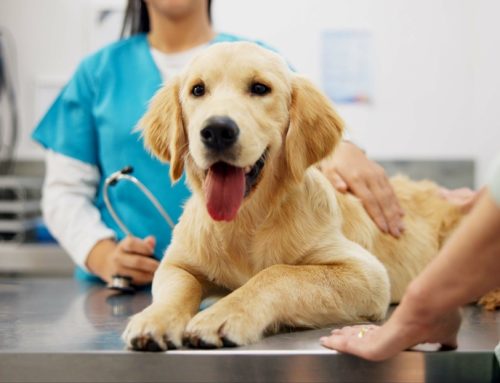
Sedation is the best way to ensure your pet’s images are diagnostic (i.e., clear and readable), but is not necessary for every situation and pet. Non-painful pets who are comfortable with handling and physical manipulation generally tolerate X-rays and ultrasound without pharmaceutical intervention.
CT is the only imaging modality we use at Central Kentucky Veterinary Center that requires full sedation.
Digital X-ray imaging for pets
X-rays are the most widely available veterinary imaging technology, although smarter, faster, and more convenient digital X-rays have now replaced the traditional, hard-copy, X-ray films.
X-ray technology uses a measured and focused radiation beam that projects an image of your pet’s internal structures onto a sensor, which sends the information to a computer. The radiation is absorbed or reflected from your pet’s soft and hard tissues, and sent to the sensor, which uses the radiation to compose a high-resolution image. X-ray radiation is reflected off hard, dense, or mineralized substances (e.g., normal bone is white or grey), passes through air (e.g., healthy lungs appear black), and is absorbed at varying levels by soft tissue (e.g., organs, cartilage, and muscle appear as grey gradients).
At Central Kentucky Veterinary Center, we most commonly use X-rays to assess the following situations:
- Emergency triage (e.g., bloat, collapsed lung, broken pelvis)
- Heart size and shape
- Lung health
- Bone fractures
- Bladder or kidney stones
- Foreign object ingestion
- Oral cavity assessment (i.e., dental X-rays)
- Orthopedic evaluation (e.g., hip or elbow dysplasia screenings, such as PennHIP and OFA, post-operative repairs)
Ultrasound technology for pets
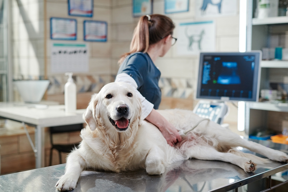
Unlike other modalities that provide a static or fixed image, ultrasound technology (i.e., sonography) uses harmless, high frequency sound waves to form a real-time and dynamic (i.e., moving) image of the pet’s internal processes, such as heart muscle contractions, intestinal motility, and mineralized crystals floating in the urinary bladder.
During an ultrasound, pets are typically placed on their back with their chest and abdomen exposed, the appropriate area is clipped and prepped with a conductive gel, and the pet is examined using a handheld transducer moved across their skin. The transducer emits painless soundwaves through the body wall and receives the returning waves that are reflected back. The reflected waves are then converted into a moving image on a computer screen that a veterinary professional observes, measures, and maps for blood flow.
Ultrasound’s versatility and minimally invasive nature make the modality popular for most soft tissue imaging needs, including:
- Heart function (i.e., echocardiogram)
- Intestinal thickness and motility
- Blood flow through the vessels
- Organ assessment (e.g., enlargement, inflammation, abnormality, tumors)
- Diagnostic sampling (e.g., needle biopsy, sterile urine or fluid collection)
- Pregnancy screening and neonate assessment
Computed tomography (CT) for pets

Central Kentucky Veterinary Center is proud to be one of the few veterinary facilities in the state to offer computed tomography (CT) imaging. With this exciting and advanced technology, we can obtain thinly sliced, cross-sectional views of bone, joints, soft tissue structures, and blood vessels in unrivaled detail.
Much like a human CT machine, our CT features a sliding gurney and a round “tube” open on both sides. Although the scanning process is relatively short (i.e., less than five minutes), CT uses multi-directional X-ray beams, so pets must be sedated or anesthetized for safety and accuracy.
CT imaging provides your pet’s veterinarian with remarkably detailed illustrations of your pet’s internal health. However, this modality is more extensive and expensive, so is generally reserved to evaluate more serious or specific conditions, such as:
- Nasal cancers or abnormalities
- Conditions affecting the chest cavity
- Airway and lung function studies
- Metastatic cancer screenings
- Congenital abnormalities (e.g., liver shunts)
- Complicated fractures
- Bone disorders or malformations (e.g., elbow dysplasia, osteochondrosis [OCD])
- Three-dimensional mapping and surgical planning
Every day at Central Kentucky Veterinary Center, our skilled and dedicated team uses advanced imaging technology on pets to go beyond the surface and provide the most accurate diagnoses. This knowledge then guides our personalized treatment plans to ensure the best possible prognosis for your pet. For more information about our advanced imaging capabilities, contact our team.


Leave A Comment